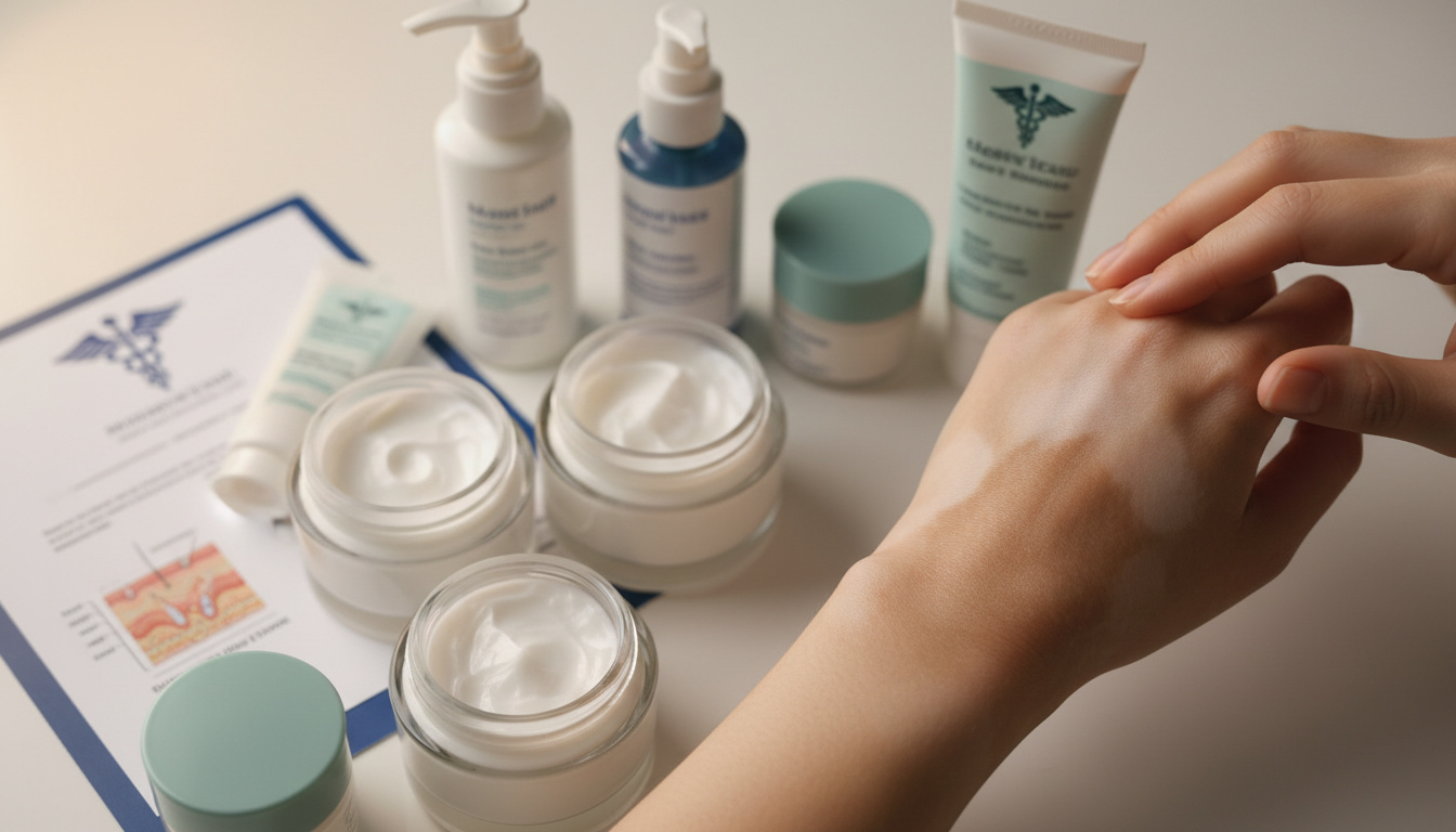
White spots emerging on arms, legs, face, or neck after sun exposure cause significant concern for millions annually. These hypopigmented patches—varying from pinprick dots to expansive patches—typically result from fungal overgrowth, autoimmune melanocyte destruction, or cumulative ultraviolet damage rather than malignancy. The contrast between tanned skin and unaffected areas often makes these spots abruptly visible during summer months, prompting immediate questions about underlying pathology.
While the majority of cases remain strictly cosmetic, distinguishing between tinea versicolor, vitiligo, pityriasis alba, and idiopathic guttate hypomelanosis proves essential for appropriate intervention. Each condition carries distinct clinical features, progression patterns, and therapeutic requirements that resist one-size-fits-all solutions.
This analysis synthesizes findings from the Cleveland Clinic, Indiana University School of Medicine dermatology researchers, and NHS dermatological guidelines to outline evidence-based identification protocols and treatment pathways.
What Causes White Spots on Skin?
Primary Triggers
Malassezia yeast overgrowth, autoimmune melanocyte targeting, post-inflammatory hypopigmentation, and sun-induced pigment cell damage represent the dominant etiologies.
Visual Indicators
Scaly, multi-colored patches suggest fungal infection; smooth, sharp-bordered patches indicate autoimmune vitiligo; tiny permanent dots signal sun damage.
Therapeutic Categories
Antifungal shampoos and creams treat tinea; topical immunosuppressants address vitiligo; moisturizers and sun protection manage environmental causes.
Professional Threshold
Dermatological consultation becomes necessary when spots spread rapidly, appear asymmetrically, or fail to respond to over-the-counter antifungal protocols.
- Tinea versicolor affects up to 50% of populations in tropical climates but remains entirely non-contagious.
- Vitiligo impacts approximately 1% of the global population, typically presenting symmetrically across both sides of the body.
- Ketoconazole 2% shampoo eliminates fungal infections in roughly 90% of cases when applied correctly over three days.
- Idiopathic guttate hypomelanosis manifests as permanent 2-5 millimeter dots exclusively on sun-exposed limbs.
- Pityriasis alba predominates among children and young adults following mild eczema episodes.
- No established causal link exists between these hypopigmentation disorders and cutaneous malignancy.
- Wood’s lamp examination immediately distinguishes fungal presentations (yellow-green fluorescence) from autoimmune vitiligo (bright white).
| Condition | Visual Presentation | Underlying Mechanism | Standard Treatment |
|---|---|---|---|
| Tinea Versicolor | Scaly, discolored patches (white, pink, red, or brown) merging unevenly | Malassezia yeast overgrowth in warm, humid conditions | Ketoconazole shampoo, terbinafine cream, selenium sulfide suspension |
| Vitiligo | Smooth, well-demarcated white patches with sharp borders | Autoimmune destruction of melanocytes | Topical calcineurin inhibitors, corticosteroids, phototherapy |
| Pityriasis Alba | Ill-defined light patches with possible fine scaling | Post-inflammatory hypopigmentation after eczema or dryness | Emollients, mild topical steroids, sun protection |
| Idiopathic Guttate Hypomelanosis | Small, round, porcelain-white macules 2-5mm in diameter | Cumulative ultraviolet damage to melanocytes | Retinoids, chemical peels, cryotherapy (limited efficacy) |
| Post-Inflammatory Hypopigmentation | Irregular light patches at sites of previous injury or inflammation | Temporary melanin production disruption | Time, topical tacrolimus, cosmetic camouflage |
| Halo Nevus | White halo surrounding existing moles | Immune response targeting nevus cells | Observation, biopsy if atypical features present |
How Do I Get Rid of White Spots on Skin?
Medical Interventions
Tinea versicolor responds robustly to topical antifungal protocols. According to GoodRX clinical data, ketoconazole 2% shampoo applied for five minutes daily over three days achieves clearance in approximately 90% of patients. Terbinafine 1% cream applied twice daily for one week shows 80% efficacy, while ciclopirox 0.77% cream requires fourteen days of twice-daily application. For widespread or recurrent infections, dermatologists prescribe oral fluconazole or itraconazole.
Vitiligo management focuses on halting progression rather than cure. Topical corticosteroids and calcineurin inhibitors (tacrolimus, pimecrolimus) serve as first-line therapies. Schweiger Dermatology Group notes that phototherapy and depigmentation procedures remain reserved for extensive cases, while skin grafting addresses stable, localized patches.
Pigment restoration following tinea versicolor treatment requires weeks to months as melanocytes repopulate treated areas. Vitiligo repigmentation occurs gradually over months, whereas idiopathic guttate hypomelanosis spots generally remain permanent despite intervention.
Home Management and Prevention
For mild tinea presentations, over-the-counter selenium sulfide or zinc pyrithione shampoos applied as body washes may reduce fungal burden. Maintaining dry skin, wearing breathable fabrics, and showering immediately after sweating limit Malassezia proliferation. West End Dermatology emphasizes that prophylactic antifungal shampoo use once monthly during summer prevents recurrence in susceptible individuals.
Vitiligo and idiopathic guttate hypomelanosis lack reliable home remedies beyond rigorous photoprotection. Broad-spectrum sunscreen prevents lesion darkening in surrounding skin, reducing contrast.
Are White Spots on Skin Dangerous?
Malignancy and Systemic Risk Assessment
None of the common hypopigmentation disorders—tinea versicolor, vitiligo, pityriasis alba, or idiopathic guttate hypomelanosis—demonstrate increased skin cancer risk or systemic complications. Research from Indiana University School of Medicine confirms zero association between these conditions and malignancy. Tinea versicolor remains superficial, never penetrating beyond the stratum corneum or spreading to internal organs.
Diagnostic Certainty and Professional Consultation
Self-diagnosis risks inappropriate treatment. Banner Health clinicians warn that visual similarity between fungal patches and early vitiligo leads to frequent misidentification. Dermatologists utilize Wood’s lamp examination—where tinea fluoresces yellow-green and vitiligo appears stark white—or microscopic examination of skin scrapings to confirm fungal presence.
Consultation becomes urgent when white spots exhibit rapid expansion, unusual border irregularity, or accompanying symptoms such as pain or persistent erythema. While exploring various health topics including Anne Hathaway (Wife of Shakespeare) – Life, Marriage and Legacy, readers should remember that historical medical understanding differs radically from modern dermatological standards.
Common Types of White Spots on Skin
Fungal Origins: Tinea Versicolor
Malassezia yeast naturally colonizes human skin, yet overgrowth triggered by heat, humidity, hormonal changes, or oily skin produces tinea versicolor. The Cleveland Clinic explains that affected areas lose pigment because the fungus produces azelaic acid, which inhibits melanin synthesis. Patches appear scaly and may present as white, pink, or brown depending on skin tone and recent sun exposure.
Tinea versicolor recurs frequently in warm climates or during summer months due to yeast reactivation. Permanent eradication proves difficult because Malassezia normally resides on skin; treatment controls rather than eliminates the organism.
Autoimmune Destruction: Vitiligo
Vitiligo involves selective autoimmune attack on melanocytes, resulting in permanent pigment loss. NHS guidelines describe smooth, milky-white patches with sharply defined borders that expand symmetrically across hands, face, and genitalia. Unlike fungal conditions, vitiligo patches show no scaling and exhibit bright white fluorescence under Wood’s lamp.
Environmental and Inflammatory Causes
Idiopathic guttate hypomelanosis presents as discrete, porcelain-white macules on sun-damaged forearms and shins, representing irreversible melanocyte loss from cumulative UV exposure. Pityriasis alba follows eczema or dermatitis episodes, creating poorly defined hypopigmented patches with subtle scaling, primarily in pediatric and adolescent populations.
Applying antifungal medications to vitiligo or idiopathic guttate hypomelanosis wastes resources and delays appropriate care. Topical steroids without diagnosis risk skin atrophy or worsening of undiagnosed fungal infections. Professional confirmation prevents these outcomes. For more information on skin conditions and their treatments, visit schweizzeit.ch.
How Do White Spots Develop Over Time?
- Initial Trigger Phase: For tinea versicolor, heat and humidity trigger yeast proliferation within days. Vitiligo may follow physical trauma or stress, though exact triggers remain unclear.
- Progression Period: Tinea patches expand and merge over weeks, becoming more visible after sun tanning. Vitiligo spreads gradually over months, with symmetrical involvement.
- Diagnostic Confirmation: Clinical examination using Wood’s lamp or microscopic scrapes establishes definitive diagnosis, typically occurring weeks to months after initial appearance.
- Treatment Implementation: Antifungal regimens require 1-3 weeks; vitiligo therapy continues for months with gradual reassessment.
- Long-term Stabilization: Tinea clears with pigment returning slowly; vitiligo may stabilize or progress unpredictably; IGH remains permanent.
What Is Definitively Known Versus Uncertain?
| Established Clinical Facts | Areas Requiring Further Study |
|---|---|
| Tinea versicolor results from Malassezia overgrowth, not hygiene deficits | Specific genetic markers predicting vitiligo progression |
| Vitiligo involves autoimmune melanocyte destruction | Individual recurrence rates for tinea in temperate climates |
| Idiopathic guttate hypomelanosis correlates with cumulative sun exposure | Prevention strategies for those with genetic susceptibility |
| Ketoconazole shampoo achieves 90% clearance in three days | Long-term safety of prolonged topical steroid use for vitiligo |
| Wood’s lamp distinguishes fungal from autoimmune hypopigmentation | Environmental triggers initiating initial vitiligo presentation |
Why Does Sun Exposure Make White Spots More Visible?
Melanin production increases in healthy skin following ultraviolet exposure, creating a tan that contrasts sharply with areas lacking functional melanocytes. In tinea versicolor, the fungus produces acids that inhibit melanin synthesis, preventing tanning in affected zones. Vitiligo patches contain no melanocytes to respond to UV stimulation. Idiopathic guttate hypomelanosis represents permanent melanocyte loss at specific sites.
This contrast effect explains why many patients first notice patches during summer or after vacation. The surrounding skin darkens while damaged areas remain pale, creating the illusion of sudden onset when the condition may have existed undetected for months. Understanding these photobiological mechanisms helps distinguish between conditions requiring antifungal intervention versus those needing immunomodulatory therapy.
Various publications explore how environmental factors influence biological processes, much like examining Juliette Lewis Movies and TV Shows – Full Filmography Guide reveals how career trajectories evolve under industry pressures.
What Do Medical Authorities Confirm?
Tinea versicolor is caused by a type of yeast called Malassezia, which naturally lives on the skin. When it grows out of control, it causes spots on the skin that are lighter or darker than surrounding skin.
Cleveland Clinic
Vitiligo is caused by the lack of a pigment called melanin in the skin. Melanin is produced by skin cells called melanocytes, and it gives your skin its colour. In vitiligo, there are not enough working melanocytes to produce enough melanin in your skin.
National Health Service
Patches in tinea versicolor feel scaly or dry and tend to merge together unevenly. Vitiligo patches are smooth with sharp borders.
Indiana University School of Medicine
What Steps Should You Take Now?
Begin with visual assessment noting texture, border definition, and distribution. If scales are present, apply over-the-counter antifungal shampoo to affected areas. For smooth, sharply bordered patches, schedule dermatological consultation immediately. Document progression through photographs to aid clinical evaluation. Maintain strict photoprotection while awaiting diagnosis. For those interested in broader cultural contexts while managing health concerns, Juliette Lewis Movies and TV Shows – Full Filmography Guide offers comprehensive entertainment coverage.
Frequently Asked Questions
Why do white spots appear after tanning?
Tanned surrounding skin darkens while affected areas cannot produce melanin. Tinea versicolor prevents tanning through fungal acids, while vitiligo and sun damage destroy or lack melanocytes entirely, creating stark contrast.
What does idiopathic guttate hypomelanosis look like?
Small, round, porcelain-white dots measuring 2-5 millimeters appear on sun-exposed arms and legs. Unlike fungal patches, these spots remain permanent, smooth, and non-scaly.
Is tinea versicolor contagious?
No. The causative Malassezia yeast naturally inhabits human skin. Overgrowth results from individual skin chemistry and environmental factors, not transmission between people.
Can vitiligo patches regain pigment naturally?
Spontaneous repigmentation occurs rarely and unpredictably. Medical treatments stimulate melanocyte migration from hair follicles, but complete restoration without intervention remains uncommon.
How quickly do antifungal treatments work?
Ketoconazole shampoo eliminates active infection in three days for most patients, though visible pigment return requires several weeks as skin cells turnover.
Are white spots always permanent?
Tinea versicolor and pityriasis alba typically resolve with treatment or time. Vitiligo and idiopathic guttate hypomelanosis generally persist indefinitely, though treatments may reduce visibility.
Should children with white facial patches see specialists?
Pityriasis alba commonly affects children and usually requires only moisturization and sun protection. However, rapid spreading or texture changes warrant pediatric dermatology evaluation.